The thorax or chest is a part of the anatomy of humans, mammals, other tetrapod animals located between the neck and the abdomen In insects, crustaceans, and the extinct trilobites, the thorax is one of the three main divisions of the creature's body, each of which is in turn composed of multiple segments The human thorax includes the thoracic cavity and the thoracic wallFeb 18, · This module of vetAnatomy is a basic atlas of normal imaging of anatomical feline radiology The 39 sampled xray images of healthy cats were performed by Susanne AEB Borofka (PhD dipl ECVDI, Utrecht, Netherland) Those images were categorized topographically into six chapters (head, vertebral column, thoracic limb, pelvic limb, thorax andThoracic radiographs should be taken at the end of inspiration (or in case of general anaesthesia during manual inflation) when lungs are fully expanded There are few exceptions, where expiratory radiographs are desirable, because increased intrathoracic pressure or contrast between gascontaining lesions and noninflated lungs help to

Radiographie Imagerie
Radio thorax chat normal
Radio thorax chat normal-Oct 15, 17 · Radiology basics of chest CT anatomy with annotated coronal images and scrollable axial images to help medical students and junior doctors learning anatomyNov 10, · Chest radiotherapy side effects Chest radiotherapy includes radiotherapy to the breast, your chest wall (if you've had surgery to remove your breast) or to your chest itself This can include radiotherapy to the lungs or to the oesophagus (your food pipe or gullet) Side effects will depend on where you're having treatment to


Imagerie Du Thorax La Radiographie A L Aide Des Affections Pulmonaires Pdf Free Download
Sep 01, 01 · (a, b) Drawings (axial view) illustrate the normal thoracic CT anatomy at the level of the T2 (a) and T3 (b) vertebral bodies (c, d) Pancoast tumor in a 62yearold man with Horner syndrome (c) Axial CT scan demonstrates a Pancoast tumor in the left upper lobe (t) The mass is contiguous with the first rib in the area of the inferior cervicalWhether thoracic radiographic findings can be used to aid clinicians in preliminarily differentiating the two tumor types before cytology or histopathology results become available Medical records, available cytologic or histologic samples, and thoracicThe axial image demonstrates that the opacity on the chest film is actually the liver As we follow the livercontour, there is this unusual shape (yellow arrow) There is discontinuity of the crus which is a nonspecific sign (small blue arrow) On the axial image there is indentation of the liver on the posterior side due to blood in the thorax
"on a ct thorax what is dependent compressive atelectasis confirmed with prone and supine imaging?Normal pressure hydrocephalus, aqueductal stenosis, Chiari I malformation Brain MRI without contrast & CSF flow study (Acqueductal stroke volume measurement) Mass MRI without and with contrast MRI contraindicated CT without and with contrast Aneurysm or AVM "Screening" MRA Head (noncontrast) @ 3T CTA head with contrastThorax Radiography Chest radiographs (CXR) are routinely abnormal and consistently demonstrate evidence of bronchopulmonary dysplasia2,6,213,218 Common features of chronic lung disease include hyperlucency, hyperinflation, persistent lung hypoplasia, decreased pulmonary vasculature, unknown opacities, persistent mediastinal shifts, and a chronic abnormal
Dr said my scan was normal" Answered by Dr Paxton Daniel Atelectasis Is lesswell inflated lung It can look like scarring orAnswer CXR Outline mediastinum What is the normal width of mediastinum at supra cardiac vessel area?You can encounter normal size heart in acute myocardial infarction or in volume overload Vascular phase This is the first phase of congestive heart failure It represents pulmonary venous hypertension Cephalization Vessels in upper chest is more prominent as a manifestation of pulmonary venous hypertension


Chat De Thorax De Rayon X Photos Libres De Droits Et Gratuites De Dreamstime


Mercredi Imagerie 2 Anorexie Amaigrissement Et Essoufflement Chez Un Rottweiler
Dec 01, 15 · Radiographic anatomy of the normal heart and the pulmonary vasculature The normal feline cardiac silhouette Thoracic radiography is one of the most commonly employed and useful tools in the diagnostic workup of cats with cardiac disease1, 2 It is used for differentiating cats with respiratory distress associated with cardiac disorders from those with respiratory(A) Dog in right lateral recumbency with thoracic limbs pulled cranially See text for anatomic boundaries of collimated thorax (B) Right lateral thoracic radiograph of dog in Figure 2A;Thoracic radiography is one of the most commonly employed diagnostic tools for the clinical evaluation of cats with suspected heart disease and is the standard diagnostic method in the confirmation of cardiogenic pulmonary edema In the past, interpretation of feline radiographs focused on a descrip


Radiographie Thoracique Images Normales Questions Et Images Fournises Par Dr Franck Durieux Dip Ecvdi Aquivet Radiographie Thoracique Images Normales Imagerie Diagnostique Et Outils Sessions Cardio Academy


Un Cas D Hypertension Arterielle Pulmonaire Chez Un Jeune Chihuahua Clinique Veterinaire Hermes Plage Et Calypso
Jan 10, 16 · Chest wall musculature and intercostal nerve (Atlas of Human Anatomy, 6th edition, Plate 177) Clinical Note Serratus anterior free flaps are often used for reconstruction of anatomic structures such as parts of the face, limbs, or diaphragmThorax thor´aks the part of the body between the neck and abdomen;A series of annotated radiographical images highlighting the key anatomical structures of the central nervous system head and neck spine thorax abdomen and pelvis upper limb lower limb Part of our Medical Imaging Anatomy Course Online


Pneumothorax Wikipedia


Radiographies Du Chat
Radio du thorax Introduction Partie 1 Docteur SynapaseRéférences 1 https//radiopaediaorg2 http//wwwradiologyassistantnlCanine Thorax Example 2 The following radiographs are the left lateral, right lateral and ventrodorsal views of the thorax of a tenyearold Mixed Breed Dog Metallic hemoclips are present in the cranial abdomen4e année médecine – Rotation 3 – 15/16 ISM Copy Module de Cardiologie Interprétation d'une radiographie thoracique Télé thorax (TLT) de face se prend en inspiration forcée, les membres supérieurs en pronation forcée les paumes en dehors Télé thorax de profil se prend le coté malade sur la plaque, les bras levés
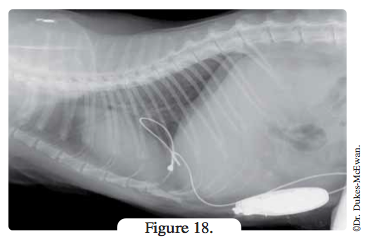


Les Arythmies Cardiaques Vetopedia Conseils Veterinaires


Le Cas De Ice Gare Aux Voitures Clinique Veterinaire Hermes Plage Et Calypso
Notice the cranial location of the thoracic limbs relative to the thoracic inlet Figure 2 (A) Dog in left lateral recumbency with thoracic limbs pulledIn summary in a normal chest Xray, anatomical borders of the peripheral bronchi are invisible However, due to pathologic changes bronchi can sometimes be distinguished When the alveoli are filled with fluid (blood, pus, mucus, edema, cells) rather than air, a density difference develops between the alveoli and bronchiNormal 2second CT anatomy of the thorax A (upper), Scan taken at hilar level Pulmonary vascular markings are shown throughout lung fields (white arrows) Descending branch of right pulmonary artery ( RP A) lies slightly lateral but predominantly anterior to air containing intermediate bronchus


Pneumothorax Et Pneumomediastins Spontanes Chez Un Chat Centre Hospitalier Clinique Veterinaire Cordeliers A Meaux 77


Radiographie Imagerie
Imaging Essentials provides comprehensive information on small animal radiography techniques The following anatomic areas have been addressed in previous columns;May 27, 16 · Cranial Lobe Vessels Assessing the absolute and relative size of pulmonary vessels is an important process of thoracic radiographic interpretation 3 The pulmonary artery and vein in the right cranial lobe are used commonly as a basis for assessing the pulmonary circulation It is important to be able to identify these two vessels specifically and to be able to compare the sizeThese articles are available at tvpjournalcom (search "Imaging Essentials") Thorax Elbow and antebrachium Abdomen Carpus and manus Pelvis Tarsus and pes Stifle joint and crus Cervical, thoracic, and lumbar spine


Radiographie Radioscopie Clinique Veterinaire Des Docteurs Martin Granel Beaufils Jumelle Calvisson


Radiographie Radioscopie Clinique Veterinaire Des Docteurs Martin Granel Beaufils Jumelle Calvisson
What is the normal width of mediastinum at tracheal level?Mediastinal abnormalities, including cardiac disease, are common causes of clinical signs related to the thorax By definition, the mediastinum is the midline potential space formed between the two pleural cavities and includes the medial portions of the right and left parietal pleura (also called the mediastinal pleural) and the space formed between these serosal membranesCalcification of the cartilage rings is a common normal finding in patients older than 40 years, particularly women, but it is seldom evident on radiographs (Fig 14) The upper limits of normal for coronal and sagittal diameters of the trachea in men are 25 and 27 mm, respectively;


Radiographies Du Chien


Radiographie Thoracique Images Anormales Session 5 Questions Et Images Fournies Par Le Dr Franck Durieux Dip Ecvdi Aquivet Radiographie Thoracique Images Anormales Imagerie Diagnostique Et Outils Sessions Cardio Academy
Abstract EVER since the introduction of the Xray in the examination of the chest radiologists have fully realized that a flat plate of the thorax is insufficient for a complete analysis of the various structures, normal and abnormal, contained in the thoracic cavityOther views (lateral or lordotic) or CT scans may be necessary In active pulmonary TB, infiltrates or consolidations and/or cavities are often seen in the upper lungs with or without mediastinal or hilar lymphadenopathy However, lesions may appear anywhere in the lungs In HIV and other immunosuppressed persons, anyIn women, they are 21 and 23 mm, respectively


Mediastin Anatomie


Mercredi Imagerie 18 Hernie Phreno Pericardique Chez Un Chat
A posterioranterior (PA) chest Xray is the standard view used;This CT scan of the upper chest (thorax) shows a malignant thyroid tumor (cancer) The dark area around the trachea (marked by the white Ushaped tip of the respiratory tube) is an area where normal tissue has been eroded and died (necrosis) as a result of tumor growthDogs with shallow, wide thoracic conformation have a short, round cardiac silhouette that, on the lateral radiograph, has a marked cranial inclination and a long area of sternal contact On the VentroDorsal or DorsoVentral views, the cardiac apex usually is located to the left of the midline and is often more difficult to identify because of


Tumeurs Pulmonaires Chez Le Chien Centre Hospitalier Veterinaire Fregis


Radiographie Du Thorax Wikipedia
Answer CXR Identify Trachea, carina, right and left main stem bronchi Why are they visible, while rest of the bronchial tree is not?The chest xray is the most frequently requested radiologic examination In fact every radiologst should be an expert in chest film reading The interpretation of a chest film requires the understanding of basic principles In this article we will focus on Normal anatomy and variants Systematic approach to the chest film using an insideoutIt is separated from the abdomen by the diaphragm Its walls are formed by the 12 pairs of ribs, attached to the sides of the spine and curving toward the front The principal organs in the thoracic cavity are the heart with its major blood vessels and the lungs with the bronchi


Pyothorax Chez Un Chat Centre Hospitalier Clinique Veterinaire Cordeliers A Meaux 77


Radiographie Boules De Fourrure
The thorax is suited well to radiographic imaging due to the inherent subject contrast afforded by the airfilled lungs A vertical xray beam configuration should be used The craniocaudal area imaged should extend from cranial to the manubrium to at least one to two vertebral body lengths caudal to the most dorsocaudal limit of the diaphragmHere is my list of normal cases for reference Thorax Normal Thorax 1 6 yearold, male neutered, canine Samoyed Normal Thorax 2 7 yearold, male neutered, canine Weimaraner Normal Thorax 3 13 yearold, female neutered, canine Siberian Husky Normal Thorax 4 13 yearold, female neutered, canine Labrador RetrieverThorax, the part of an animal's body between its head and its midsection In vertebrates (fishes, amphibians, reptiles, birds, and mammals), the thorax is the chest, with the chest being that part of the body between the neck and the abdomen The vertebrate thorax contains the chief organs of


Chylothorax Chez Le Chat Centre Hospitalier Veterinaire Fregis Arcueil


Diagnostic D Autres Pathologies Des Poumons
ObjectiveTo evaluate the severity and extent of lung disease using thoracic computed radiography (CR) compared to contrastenhanced multidetector computed tomography (MDCT) of the thorax in calves with naturally occurring respiratory disease and to evaluate the feasibility and safety of performing contrastenhanced thoracic multidetector MDCT examinations in sedated calves(Slideset Comment examiner une radio du thorax ?) slide 15 Lorsque plusieurs tubulures centrales se chevauchent, il peut être plus difficile de déterminer leur emplacement Ce cliché d'une radiographie thoracique portative AP montre une tubulure dans la veine jugulaire interne droite et une tubulure dans la veine sous clavière droiteThe normal thorax is well suited to radiographic evaluation because there is marked inherent contrast between the airfilled, fluidfilled, soft tissue, and bony structures that comprise the thoracic viscera and thoracic wall As has been stated before, at least 2 orthogonal views of the thorax are required for complete and accurate interpretation For routine evaluation of the thorax


Pyothorax Chez Un Chat Centre Hospitalier Clinique Veterinaire Cordeliers A Meaux 77


Clinique Veterinaire Des Bourgeolles Donzenac Brive Deroulement Radiographie Chien Chat Radiographie Veterinaire
Figure Chest xray of a patient with pneumonia A, Posteroanterior view reveals a patchy opacity overlying the left lung base (large arrow)Note that the left cardiac border is preserved, indicating that the pneumonia is more likely in the left lower lobe If it were lingular and abutting the left cardiac border, the left cardiac border would be obscured (the "silhouette" sign)


Blogue Animages Pour Vous Guider Dans Vos Diagnostics Page 30


Radiographie Thoracique Images Normales Questions Et Images Fournises Par Dr Franck Durieux Dip Ecvdi Aquivet Radiographie Thoracique Images Normales Imagerie Diagnostique Et Outils Sessions Cardio Academy


Pyothorax Chez Un Chat Centre Hospitalier Clinique Veterinaire Cordeliers A Meaux 77


Radiographie Imagerie


Maladies Chez Le Furet Club Francais Des Amateurs Du Furet
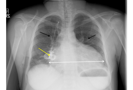


Comment Examiner Une Radio Du Thorax Medscape


Diagnostic D Autres Pathologies Des Poumons
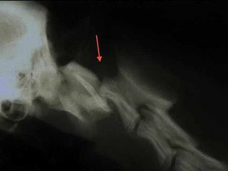


Radiographie


Clinique Veterinaire Des Bourgeolles Donzenac Brive Deroulement Radiographie Chien Chat Radiographie Veterinaire
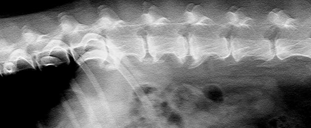


Radiographie
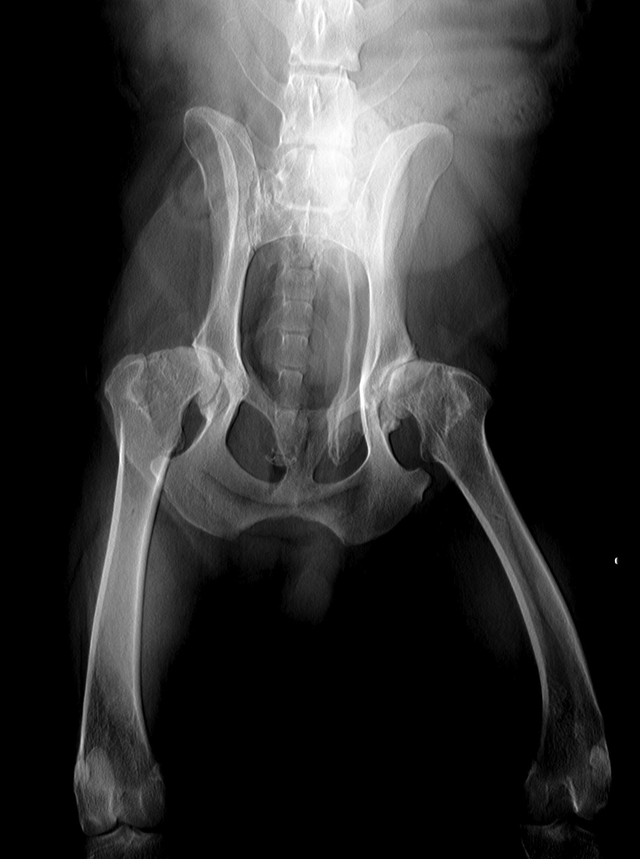


Radiographie


Radiographie Thoracique Images Normales Questions Et Images Fournises Par Dr Franck Durieux Dip Ecvdi Aquivet Radiographie Thoracique Images Normales Imagerie Diagnostique Et Outils Sessions Cardio Academy


Chat De Thorax De Rayon X Photos Libres De Droits Et Gratuites De Dreamstime
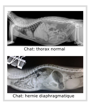


Blessures Chez Les Petits Animaux


Que Peut On Voir Ne Peut On Pas Voir A L Examen Radiographique Clinique Veterinaire Mairie D Issy


Radiographie Clinique Veterinaire Saint Francois


Chat De Thorax De Rayon X Photos Libres De Droits Et Gratuites De Dreamstime


Radiographies Du Chat


Diagnostic Et Traitement D Un Pectus Excavatum Chez Un Chat Chvsm


Radiographie Du Thorax Normale 1 Youtube


Que Peut On Voir Ne Peut On Pas Voir A L Examen Radiographique Clinique Veterinaire Mairie D Issy


Radiographie Imagerie
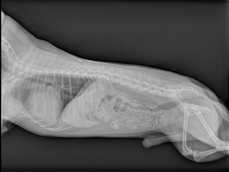


Les Maladies Cardiaques Chez Le Chien Clinique Veterinaire Du Boulingrin A Rouen


Chat De Thorax De Rayon X Photos Libres De Droits Et Gratuites De Dreamstime


Diagnostic Et Traitement D Un Pectus Excavatum Chez Un Chat Chvsm


Dyspnee Chez Un Chat


Cardiomyopathie Hypertrophique Cmh Chez Le Chat Centre Hospitalier Veterinaire Fregis


Radiographies Du Chat
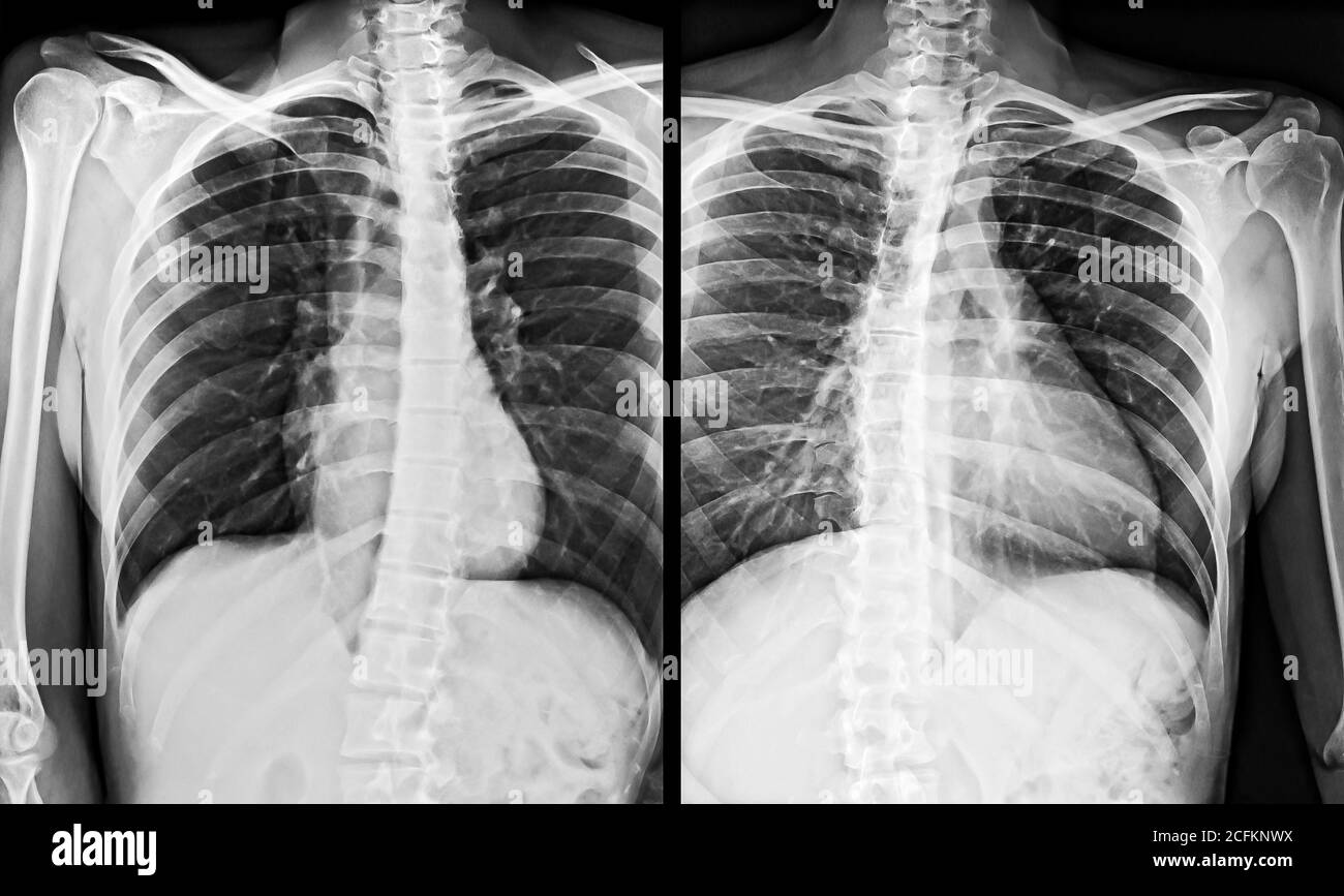


Chest Thorax X Ray Banque D Image Et Photos Alamy


Pdf Traitement Du Pectus Excavatum Chez Le Chat


Radiographie Radioscopie Clinique Veterinaire Des Docteurs Martin Granel Beaufils Jumelle Calvisson


Radiographie Boules De Fourrure


Radiographie Radioscopie Clinique Veterinaire Des Docteurs Martin Granel Beaufils Jumelle Calvisson


Dyspnee Chez Un Chat


Radiographie Radioscopie Clinique Veterinaire Des Docteurs Martin Granel Beaufils Jumelle Calvisson


Clinique Veterinaire Du Dr Lustman A Saint Denis Conseils Archives Page 7 Sur 10 Clinique Veterinaire Du Dr Lustman A Saint Denis
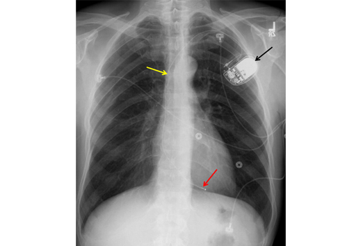


Comment Examiner Une Radio Du Thorax Medscape


Imagerie Du Thorax La Radiographie A L Aide Des Affections Pulmonaires Pdf Free Download


Radiographie Radioscopie Clinique Veterinaire Des Docteurs Martin Granel Beaufils Jumelle Calvisson


Cardiologie Clinique Veterinaire Des Docteurs Martin Granel Beaufils Jumelle Calvisson


Imagerie Du Thorax La Radiographie A L Aide Des Affections Pulmonaires Pdf Free Download
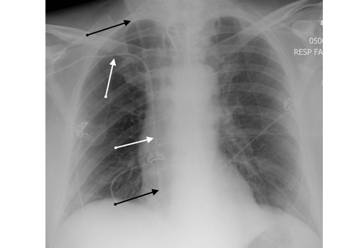


Comment Examiner Une Radio Du Thorax Medscape


File Radiographie Thoracique Chez Un Chat Jpg Wikimedia Commons


Radiographie Radioscopie Clinique Veterinaire Des Docteurs Martin Granel Beaufils Jumelle Calvisson
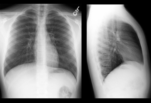


Comment Examiner Une Radio Du Thorax Medscape


Diagnostic Et Traitement D Un Pectus Excavatum Chez Un Chat Chvsm


œdeme Pulmonaire Cardiogenique Du Chat
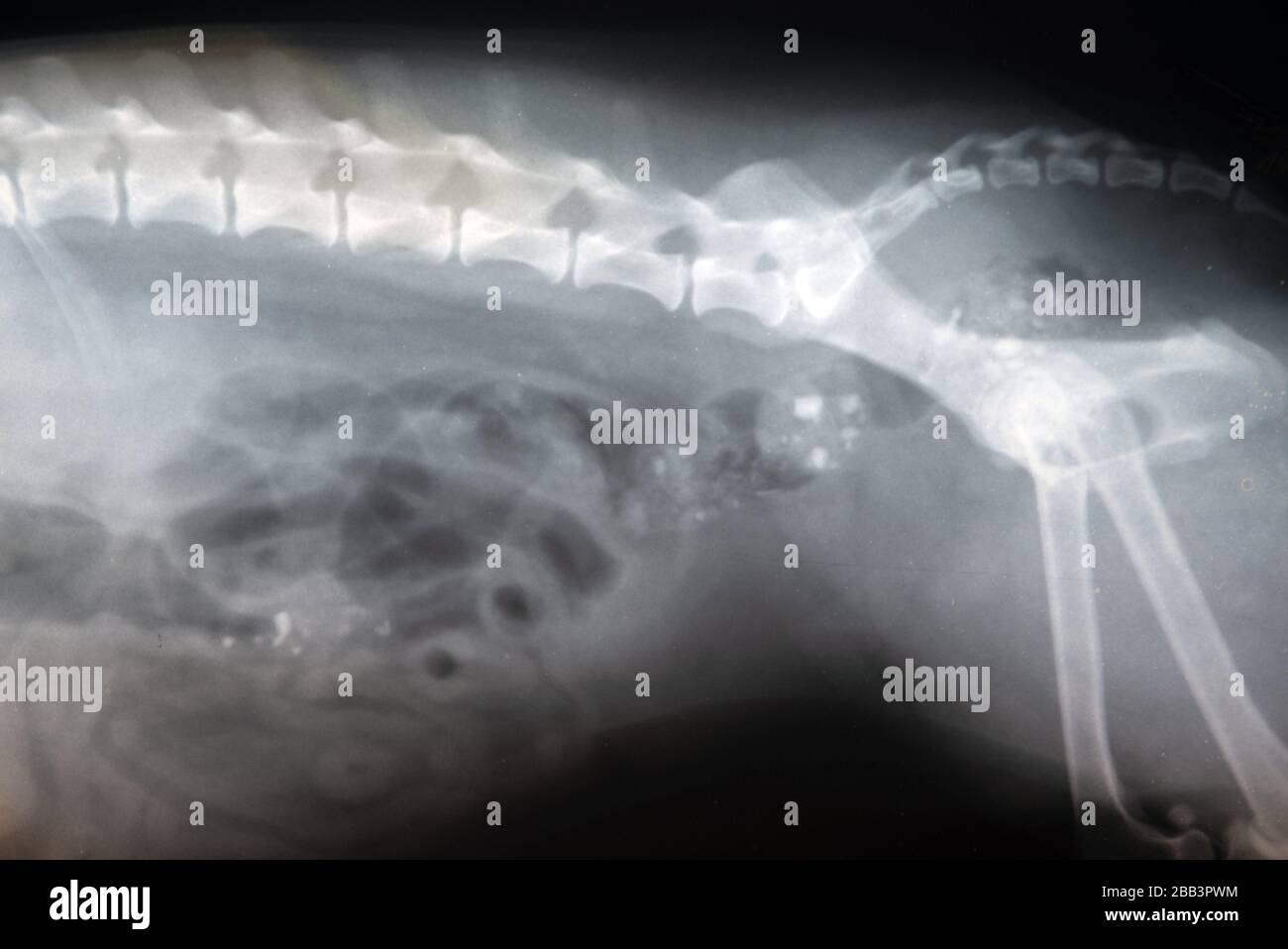


Thorax Anatomy Banque D Image Et Photos Alamy


Radiographie Du Thorax Anatomie


Adenocarcinome Pulmonaire Chez Une Jeune Chatte Centre Hospitalier Clinique Veterinaire Cordeliers A Meaux 77


Radiographies Du Chien


Adenocarcinome Pulmonaire Chez Une Jeune Chatte Centre Hospitalier Clinique Veterinaire Cordeliers A Meaux 77


Les Tumeurs Chez Le Chat Chat Fait Du Bien
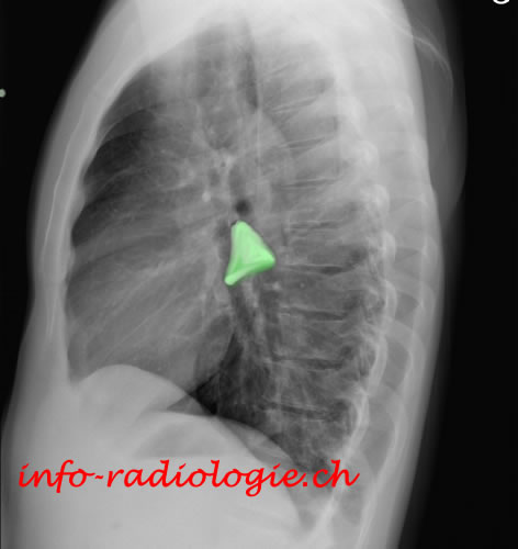


Radiographie Du Thorax Anatomie


Chat De Thorax De Rayon X Photos Libres De Droits Et Gratuites De Dreamstime


Un Chat Blesse Par Balle Ses Proprietaires Portent Plainte Ladepeche Fr


Que Peut On Voir Ne Peut On Pas Voir A L Examen Radiographique Clinique Veterinaire Mairie D Issy


Radiographie Boules De Fourrure


œdeme Pulmonaire Cardiogenique Du Chat


Radio Du Thorax Introduction Partie 1 Docteur Synapse Youtube


Imagerie Du Foie Et Du Pancreas Royal Canin


Pneumothorax Et Pneumomediastins Spontanes Chez Un Chat Centre Hospitalier Clinique Veterinaire Cordeliers A Meaux 77


Radiographie Radioscopie Clinique Veterinaire Des Docteurs Martin Granel Beaufils Jumelle Calvisson


Radiographies Du Thorax Troubles Cardiaques Et Vasculaires Manuels Msd Pour Le Grand Public


Aucun commentaire:
Publier un commentaire